Videos
| Videos | ||
|---|---|---|
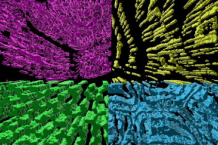
A fly-through rendering of the mitochondrial network within an oxidative mouse skeletal muscle cell. Mitochondria are colored according to their location relative to the adjacent contractile structures. | Fly-through rendering of the interior structures of a newborn mouse skeletal muscle highlighting cell and organelle membrane as well as myosin filaments. | |
3D rendering of the interior of a mouse retina mitochondrion. Outer membrane (transparent), inner boundary membrane (transparent), inner membrane cristae (magenta), matrix (yellow), and nucleoids (blue) are shown. | 3D rendering of the Z-disk sheets (light green) within the jump muscle of the fruit fly, Drosophila melanogaster. Individual z-disks (various colors) and contractile sarcomeres (magenta) are also shown. | |
3D rendering of the branching contractile structures within a newborn mouse skeletal muscle. Each color represents an individual myofibril segment. The cell membrane (green) and a nucleus (cyan) are also shown. | 3D rendering of the interior of a newborn mouse skeletal muscle. Mitochondria (red), sarcotubular network (green), lipid droplets (yellow), cell membranes (white), and myosin filaments (blue) are shown. | |
Meet the Team

Brian Glancy, Ph.D.
Brian Glancy graduated with a B.A. in Sport Science from the University of the Pacific prior to receiving a Master’s degree in Kinesiology and a Ph.D. in Exercise Science from Arizona State University working with Wayne Willis. He was a postdoctoral fellow with Robert Balaban at the National Heart, Lung, and Blood Institute from 2009 to 2016. Dr. Glancy became an Earl Stadtman Investigator at the NIH with a dual appointment between NHLBI and NIAMS in 2016 and became a tenured Senior Investigator in 2023. He is a member of the American College of Sports Medicine and the American Physiological Society.
Contact the lab

Yingfan Zhang, Ph.D.

Prasanna Katti, Ph.D.

Hailey Parry, Ph.D.
Christian Dang


