Senior Investigator Research Interests
Skeletal muscle is the most abundant tissue in humans and faces near instantaneous changes in demand for force production lasting from seconds to minutes to hours. Initiating and maintaining muscle contraction requires rapid, coordinated movement of signals and material within and among various structures located throughout the relatively large muscle cell. The Muscle Energetics Laboratory focuses on the energy distribution aspect of continued muscle contraction, deficits in which have been implicated in many pathologies including diabetes, cardiovascular disease, muscular dystrophy, and aging. In particular, we aim to determine how mitochondrial and contractile networks are optimized as part of the integrated muscle cell to maintain energy homeostasis during the large change in energy demand caused by the onset of muscle contraction. Ongoing efforts are centered around the structure, function, composition, and developmental regulation of mitochondrial and contractile networks with the goal of gaining better control of and understanding the functional consequences of altering spatial relationships within the muscle energy distribution system.

|

|

|
Videos
| Videos | ||
|---|---|---|
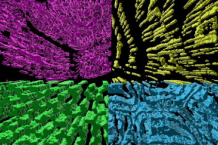
A fly-through rendering of the mitochondrial network within an oxidative mouse skeletal muscle cell. Mitochondria are colored according to their location relative to the adjacent contractile structures. | Fly-through rendering of the interior structures of a newborn mouse skeletal muscle highlighting cell and organelle membrane as well as myosin filaments. | |
3D rendering of the interior of a mouse retina mitochondrion. Outer membrane (transparent), inner boundary membrane (transparent), inner membrane cristae (magenta), matrix (yellow), and nucleoids (blue) are shown. | 3D rendering of the Z-disk sheets (light green) within the jump muscle of the fruit fly, Drosophila melanogaster. Individual z-disks (various colors) and contractile sarcomeres (magenta) are also shown. | |
3D rendering of the branching contractile structures within a newborn mouse skeletal muscle. Each color represents an individual myofibril segment. The cell membrane (green) and a nucleus (cyan) are also shown. | 3D rendering of the interior of a newborn mouse skeletal muscle. Mitochondria (red), sarcotubular network (green), lipid droplets (yellow), cell membranes (white), and myosin filaments (blue) are shown. | |
Meet the Team

Brian Glancy, Ph.D.
Brian Glancy graduated with a B.A. in Sport Science from the University of the Pacific prior to receiving a Master’s degree in Kinesiology and a Ph.D. in Exercise Science from Arizona State University working with Wayne Willis. He was a postdoctoral fellow with Robert Balaban at the National Heart, Lung, and Blood Institute from 2009 to 2016. Dr. Glancy became an Earl Stadtman Investigator at the NIH with a dual appointment between NHLBI and NIAMS in 2016 and became a tenured Senior Investigator in 2023. He is a member of the American College of Sports Medicine and the American Physiological Society.
Contact the lab

Yingfan Zhang, Ph.D.

Prasanna Katti, Ph.D.

Hailey Parry, Ph.D.
Christian Dang


