MORE INFORMATION
How the Heart Works What the Heart Looks Like
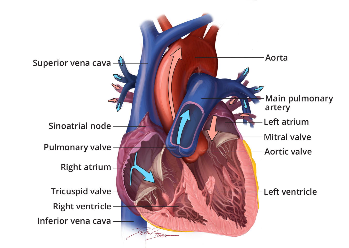
Your heart is in the center of your chest, near your lungs. It has four hollow chambers surrounded by muscle and other heart tissue. The chambers are separated by heart valves, which make sure that the blood keeps flowing in the right direction. Read more about heart valves and how they help blood flow through the heart.

Heart chambers
The two upper chambers of your heart are called , and the two lower chambers are called . Blood flows from the body and lungs to the atria and from the atria to the ventricles. The ventricles pump blood out of the heart to the lungs and other parts of the body. An internal wall of tissue divides the right and left sides of your heart. This wall is called the septum.
Heart tissue
The heart is made of three layers of tissue.
- Endocardium is the thin inner lining of the heart chambers and also forms the surface of the valves.
- Myocardium is the thick middle layer of muscle that allows your heart chambers to contract and relax to pump blood to your body.
- Pericardium is the sac that surrounds your heart. Made of thin layers of tissue, it holds the heart in place and protects it. A small amount of fluid between the layers helps reduce friction between the beating heart and surrounding tissues.
Some conditions can affect the heart's tissue.
- Cardiomyopathy is when the heart muscle becomes enlarged, thick, or rigid. As cardiomyopathy worsens, the heart becomes weaker and is less able to pump blood through the body and maintain a normal electrical rhythm.
- Heart inflammation is in one or more of the layers of tissue in the heart, including the pericardium, myocardium, or endocardium. This can lead to serious complications, including heart failure, cardiogenic shock, or irregular heart rhythm.
- Congenital heart disease is when the heart does not develop in the typical way. A heart defect can happen at any point during development of an unborn baby, or embryo, inside the pregnant mother.

