Laboratory of Imaging Technology

The Laboratory of Imaging Technology Program, led by Adrienne Campbell-Washburn, is focused on the development of advanced MRI techniques that leverage modern acquisition and reconstruction techniques for cardiac imaging, lung imaging and MRI-guided cardiovascular catheterization procedures.

Adrienne

Videos

Videos
Multi-section imaging of the lung using T2-weighted MRI (1.1mm x 1.1 mm x 6mm) in a woman with lymphangioleiomyomatosis resulting in innumerable thin walled pulmonary cysts. Adapted from Campbell-Washburn AE, et al, Radiology: Cardiothoracic Imaging https://doi.org/10.1148/ryct.2021200611 (Online ahead of print)
ECG-gated spiral in-out bSSFP cine imaging at 0.55T (spiral readout duration = 6.5ms, TR = 8ms). Advanced acquisition strategies increase SNR with limited artifacts at 0.55T. Adapted from Restivo MC, et al, Magn Reson Med, 2020; 84(5):2364-2375.
Interactive real-time imaging for MRI-guided cardiovascular catheterization at 0.55T. The bright gadolinium-filled balloon at the tip of the catheter is navigated in different heart chambers. Adapted from Campbell-Washburn et al, Radiology, 2019; 293(2):384-393.

Images

Late gadolinium enhancement (scar imaging) at 0.55T and 1.5T in three example patients with myocardial infarction.
Click the image to expand

Late gadolinium enhancement (scar imaging) at 0.55T and 1.5T in three example patients with myocardial infarction.

These comparisons demonstrate diagnostic similarity between the research 0.55T MRI and clinical 1.5T MRI. Adapted from Bandettini et al, JACC: Cardiovascular Imaging, DOI: 10.1016/j.jcmg.2021.02.024 (Online ahead of print)

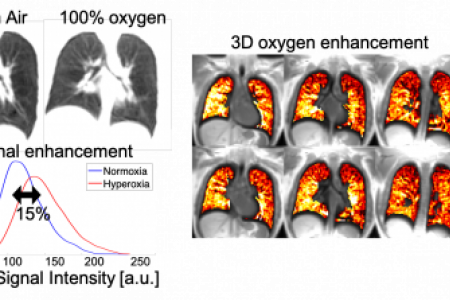
Measurement of regional lung function using oxygen-enhanced MRI.
Click the image to expand

Measurement of regional lung function using oxygen-enhanced MRI.

3D stack-of-spirals ultrashort-TE imaging in a healthy volunteer during inhalation of room air versus 100% oxygen. Lung signal intensity histograms demonstrate signal enhancement on T1-weighted imaging with hyperoxia, which results in signal enhancement visible in percent enhancement maps. Adapted from Bhattacharya et al, NMR in Biomedicine, 2021;e4562 (Online ahead of print)

Clinical Trials and Studies

RECLUTAMIENTO
Adultos, Ancianos
All
Se Aceptan Voluntaries Saludables
Desarrollo de nuevas técnicas de imagen por resonancia magnética cardiovascular. Este estudio compara la eficacia de dos tipos de escáneres de resonancia magnética (RM) para detectar diferentes tipos de cardiopatías, como la cardiopatía coronaria, la insuficiencia cardíaca, las cardiopatías congénitas y las valvulopatías cardíacas. El nuevo tipo de escáner de RM de este estudio usa menos energía que un escáner tradicional y puede ser adecuado para personas que tienen dispositivos metálicos en el cuerpo. Para participar en este estudio, debe ser sano o debe tener diagnóstico de cardiopatía coronaria estable y debe tener al menos 18 años. Este estudio se lleva a cabo en Bethesda (Maryland).

Meet the Team

Adrienne

Adrienne Campbell-Washburn, Ph.D.

Senior Investigator​ ​

Dr. Adrienne Campbell-Washburn graduated with her B.Sc. in physics from the University of Western Ontario (Canada), and received her PhD in Medical Physics from University College London (UK). She joined NHLBI in 2013 as a postdoctoral fellow in the Laboratory of Cardiovascular Interventions. Dr. Campbell-Washburn was appointed Staff Scientist in 2016 and director of the MR Technology Program in 2017. As of 2020, Dr. Campbell-Washburn became an Earl Stadtman Investigator in NHLBI and the chief of the MRI Technology Program. Dr. Campbell-Washburn is a Junior Fellow of the International Society for Magnetic Resonance in Medicine, a member of the Society for Cardiovascular MRI, and member of the Magnetic Resonance in Medicine editorial board. Her lab has pioneered 0.55T MRI technology for imaging the heart and lungs.

Alumni

Burcu Basar, MSc

Engineer

Björn Wieslander, M.D., Ph.D.

Adv. Pulmonary Imaging Fellow